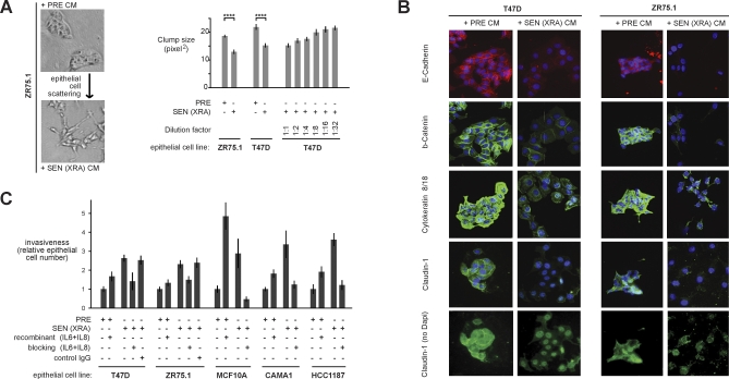
Figure 4. Novel SASP Biological Activities and Key Factors.
(A) T47D and ZR75.1 cells were incubated for 2 d with CM from PRE fibroblasts, or SEN fibroblasts induced by XRA. The cells were photographed under phase contrast, or analyzed for cluster size using an automated Cellomix imager and software. Smaller cluster or clump sizes (pixel2) indicate greater scattering. The senescence inducer is given in parentheses. Quadruple asterisks (****) indicate p < 0.001. Error bars indicate the standard deviation around the mean.
(B) T47D and ZR75.1 cells were incubated with the indicated CM for 3 d and immunostained for the indicated EMT marker proteins. Induction of the mesenchymal marker vimentin by SEN (XRA) CM is shown by the western blot in Figure 7A.
(C) Epithelial cell invasion was measured using Boyden chambers containing CM alone or CM plus IL-6 and IL-8 recombinant proteins or IL-6 and IL-8 blocking antibodies, as described in Materials and Methods. After 16–24 h, invasion was scored by counting the number of cells on the underside of the membrane. Invasion stimulated by PRE CM was given a value of one, and other conditions were normalized to this value. Invasion was significantly stimulated by recombinant protein and significantly inhibited by antibodies (p < 0.05). Error bars indicate the standard deviation around the mean.

