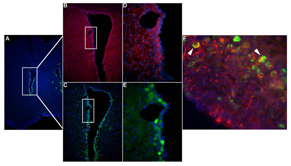
Figure 1. MCMV-infection of endogenous NSCs in vivo.
Coronal murine brain sections immunostained with anti-β-galactosidase antibody display staining indicative of viral infection with the Lac Z-containing recombinant MCMV RM461. (A & C) Lower magnifications demonstrate that MCMV is localized to cells surrounding the ventricles. (E) Higher power micrographs demonstrate that the infection occurs in periventricular cells (green cells). (B & D) Adjacent serial sections, immunostained to detect the stem cell marker nestin, show that NSCs are co-localized to the infected region (red cells). (F) Double-immunohistochemical staining for β-galactosidase and nestin reveals that MCMV infects NSCs in vivo (white arrows point to dual positive yellow cells).

