
Fig. 3.
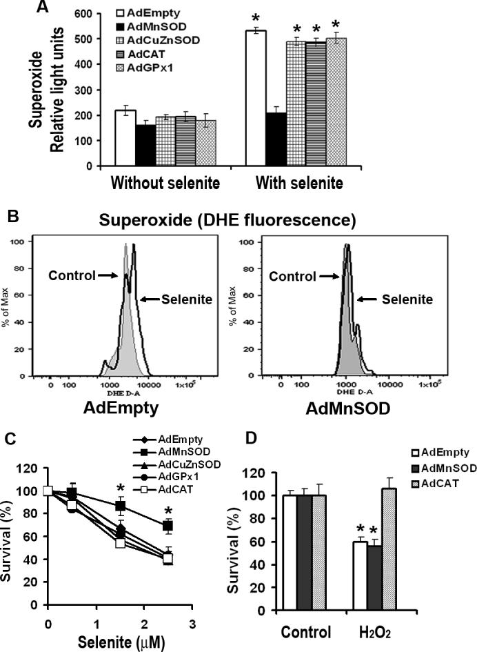
Effect of overexpression of AEs on selenite-induced production of superoxide and viability in LNCaP cells. a Levels of selenite-induced superoxide production in LNCaP cells with overexpression of MnSOD, CuZnSOD, CAT, or GPx1. Cells were pretreated with 2.5 μM selenite for 6 min and then harvested for chemiluminescence assay using a luminometer after transduction with 50 MOI AdEmpty, AdMnSOD, AdCuZnSOD, AdCAT, or AdGPx1 for 48 h. The data were obtained from three independent experiments and shown as means ± SD. *P < 0.05 compared with control cells without selenite treatment. b Levels of superoxide in cells transduced with AdEmpty or AdMnSOD. After transduction with 50 MOI AdEmpty or AdMnSOD for 48 h, cells were harvested in suspension and then incubated with 2.5 μM selenite and 5 μM DHE for 5 min. DHE fluorescence was detected using a flow cytometer. c MTT assay of cell viability. Cells were plated in 96-well plates overnight, transduced with 50 MOI AdEmpty, AdMnSOD, AdCuZnSOD, AdCAT, or AdGPx1 for 48 h, and then treated with different concentrations of selenite for an additional 5 days. Data are presented as means ± SD of three independent experiments. * P < 0.05 compared with AdEmpty at the corresponding concentrations of selenite. d MTT assay of cell viability. Cells were grown in 96-well plates overnight, transduced with 50 MOI AdEmpty, AdMnSOD, or AdCAT for 48 h, and then treated with 50 μM H2O2 for 24 h. Data are presented as means ± SD of three independent experiments. * P < 0.05 compared with control cells without H2O2 treatment.