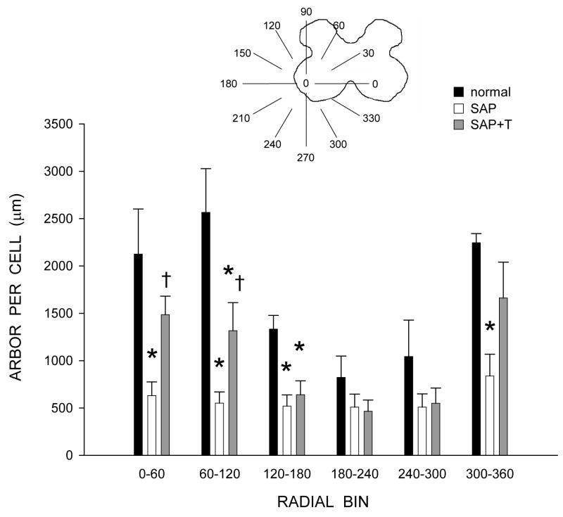
Figure 5.
Inset: Drawing of spinal gray matter divided into radial sectors for measure of quadriceps motoneuron dendritic distribution. Length per radial bin of quadriceps dendrites of normal males (black bars) and saporin-injected animals that were either untreated (SAP, unfilled bars), or treated with testosterone (SAP+T, gray bars). For graphic purposes, dendritic length measures have been collapsed into 6 bins of 60° each. Quadriceps motoneuron dendritic arbors display a non-uniform distribution, with the majority of the arbor located between 300° and 120°. Following saporin-induced motoneuron death, surviving nearby motoneurons had reduced dendritic length in every radial bin. Treatment with testosterone attenuated this reduction. Bar heights represent means ± SEM. * indicates significantly different from normal males. † indicates significantly different from untreated saporin-injected animals.

