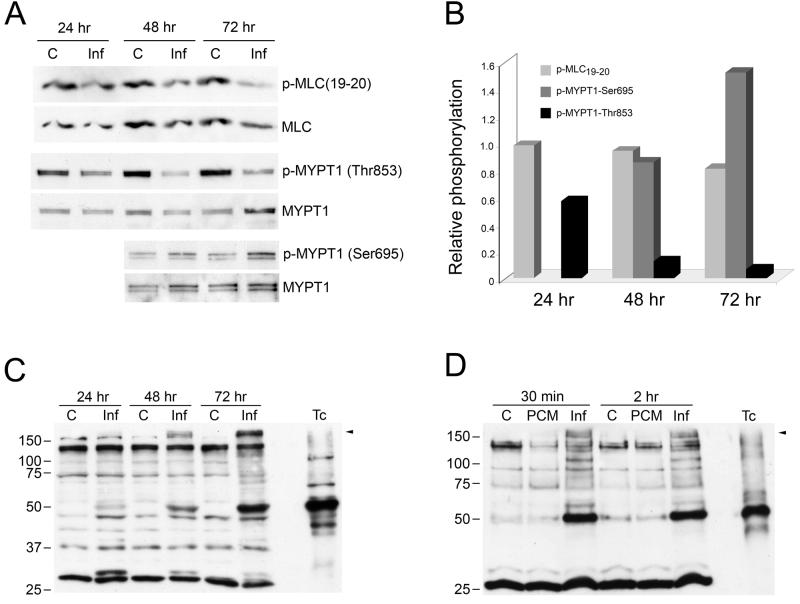
Figure 6. T. cruzi elicits changes in Rho kinase and PKA-dependent substrate phosphorylation.
Lysates prepared from mock- or T. cruzi-infected HFF (C or Inf) were blotted and probed with antibodies to phospho-MLC(19-20), phospho-MYPT1-Thr853, phospho-MYPT1-Ser695, stripped and reprobed with MLC or MYPT1 as appropriate (A) and densitometric analysis of the bands was carried out to determine the average relative phosphorylation levels from 2 independent experiments (B). Mock- and T. cruzi-infected (C; Inf) or PCM-treated (PCM) HFF lysates were subjected to western blotting and probed with antiphospho-PKA substrate antibody (C,D). A mixture of isolated parasites 2 × 10⁶trypomastigote/amastigote mix was included as a control for phosphorylated parasite proteins (Tc). Arrow indicates differentially phosphorylated protein band that is not present in isolated parasites.

