
Figure 2. Electron micrographs of atypically large nonperforated synapses.
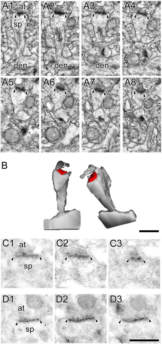
A: Serial sections through an atypically large nonperforated (ANP) synapse (arrowheads) between an axon terminal (at) and a spine (sp), which is seen connecting to its parent dendrite (den). A spine apparatus is observed in the spine head/neck region. B: 3-dimensional reconstructions of the ANP synapse and its parent spine in their original orientation (Left) and rotated (Right) to illustrate the continuous shape and large size of its postsynaptic density (PSD). Note the nonsynaptic spinule emanating from a perisynaptic region of the spine head. C, D: Serial sections through ANP synapses between axon terminals (at) and spines (sp), which were always immunopositive for AMPA-type receptors (C) and NMDA-type receptors (D). Scale bars = 0.5 μm. Though the ANP synapses showing AMPA-type and NMDA-type are different synapses, all ANP synapses were immunopositive for both. It is likely, then, that each of these synapses, had they been immunostained for both AMPA-type and NMDA-type receptors, would be immunopositive for both. Also note that all but 1 of the immunogold particles in panel C1 are considered synaptic according to our criteria (i.e., on or otherwise within 20 nm of the postsynaptic density, or in the synaptic cleft).