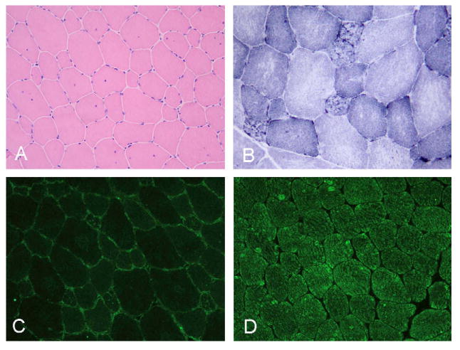
Fig. 3.

Muscle biopsy showing (A) wide variation in the fiber size with internal nuclei and mild endomysial fibrosis. On NADH reaction with (B) most of the atrophic fibers are lobulated. Lack of telethonin (C) as compared with normal sarcomeric labeling in the control (D). A, C and D 200×; B 400×.
