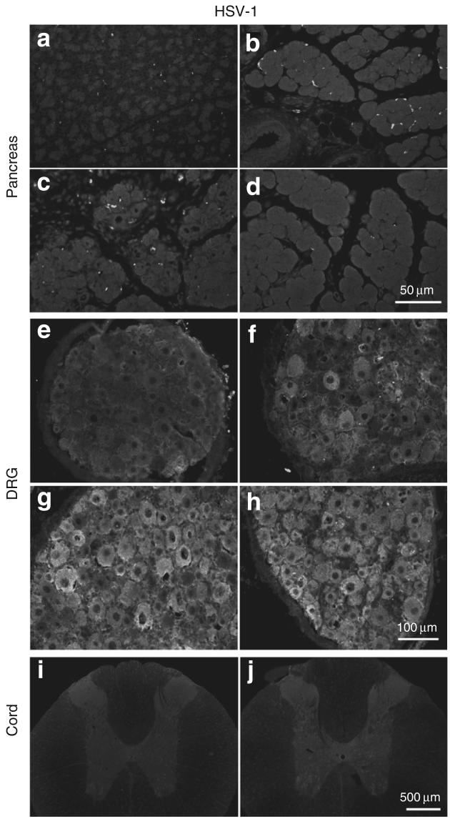
Figure 3. Photomicrographs of immunohistochemical staining of HSV-1 proteins in pancreas.
HSV-1 is shown in (a-d), dorsol root ganglia (DRG, e-h) and spinal cord (i, j) on day 7 of DBTC-induced pancreatitis or in controls. Pancreas after (a) sham surgery and after DBTC-induced pancreatitis with (b) vehicle. (c) HSV-β-gal. (d) HSV-ENK. (e) DRG from a rat after sham surgery. DRG from rats with DBTC-induced pancreatitis after application of (f) vehicle. (g) HSV-β-gal. (h) HSV-ENK. (i) Spinal cord from a rat after sham surgery. (j) Spinal cord from a rat with DBTC pancreatitis after application of HSV-ENK. ENK, enkephalin.

