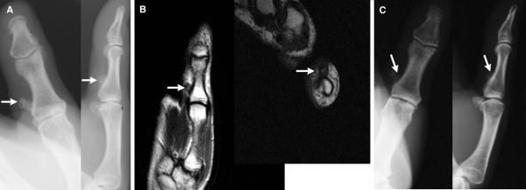
FIG. 1. Imaging results for a 51-year-old woman with bizarre parosteal osteochondromatous proliferation at the proximal phalanx of her left thumb. (A) Anteroposterior and lateral radiographs showing a calcified mass (arrow) on the left thumb attached to the proximal phalanx without alteration of the underlying cortex. (B) Τ1-weighted magnetic resonance images (sagittal, axial) showing a low-signal lesion (arrow) extending from the thumb. There is normal signal intensity of the cortex and the bone marrow of the underlying bone. (C) Anteroposterior and lateral radiographs of an asymptomatic, stable local recurrence (arrow) 16 months after excision.

An official website of the United States government
Here's how you know
Official websites use .gov
A
.gov website belongs to an official
government organization in the United States.
Secure .gov websites use HTTPS
A lock (
) or https:// means you've safely
connected to the .gov website. Share sensitive
information only on official, secure websites.
