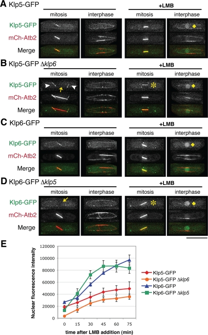
Figure 1.
Klp5 and Klp6 are interdependent for nuclear mitotic localization. Wild-type (A and C), Δklp6 (B), or Δklp5 (D) strains containing mCherry-Atb2 and either Klp5-GFP (A and B) or Klp6-GFP (C and D) were used to visualize microtubules and Klp5/6 localization. (A–D) Representative images before (first and second panels) or after LMB addition (45 min; third and fourth panels) are shown during mitosis (first and third panels) or interphase cells (second and fourth panels). Cytoplasmic astral microtubules and dim nuclei are marked with arrowheads and arrows, respectively. After LMB addition, both Klp5 and Klp6 accumulate in the nucleoplasm (B and D; diamonds) or localize to nuclear spindles (asterisks) in the absence of each counterpart. In merged images, GFP-Klp5/6 and mCherry-Atb2 are shown in green and red, respectively. Bar, 10 μm. (E) Kinetics of nuclear accumulation during interphase after addition of LMB (0–75 min) are plotted in individual strains shown in A–D. n ≥5 for each strain.

