Figure 2.
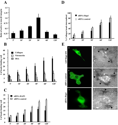
(A) For microscopic observations, cells were incubated with collagen-coated beads for different lengths of time, fixed, and stained for activated β1-integrin (mAb; clone 9EG7) and total β1-integrin (KMI6). The histogram shows the ratio of fluorescence intensities around the beads of active β1-integrin to total β1-integrin (mean ± SEM). (B) Collagen-coated beads showed progressive increases in binding over 120 min compared with vitronectin or BSA-coated beads. (C and D) Rap1-deficient cells exhibit reduced collagen-bead binding and bead internalization compared with cells treated with control siRNA. (E) Collagen uptake in cells expressing GFP-Rap1 DN (a and b), and transfected with fluorescently labeled control (c and d) or c Rap1 (e and f) siRNA after 4 h. Cells treated with control siRNA show substantial clearance of fluorescent collagen (black arrows in d) compared with Rap1 knockdown cells (f) or cells expressing dominant negative (DN) Rap1 (b). (A–D) The data represent mean ± SEM for three independent experiments, 25 cells per experiment.

