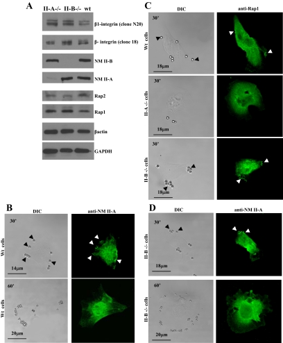
Figure 6.
(A) Total cell lysates from NM II-A null, NM II-B null, and wild-type (Wt) ES cells were immunoblotted for the indicated proteins. (B) Immunostained WT cells show accumulation of NM II-A in phagocytic cups at bead-binding sites at 30 min. By 60 min, NM II-A staining at bead sites disappeared. (C) WT cells and NM II-B null cells showed accumulation of Rap1 at bead sites after 30-min incubation, but the absence of NM II-A prevented localization of Rap1 to beads. (D) In NM II-B null cells, NM II-A localizes to beads at 30 min and is absent at 60 min.

