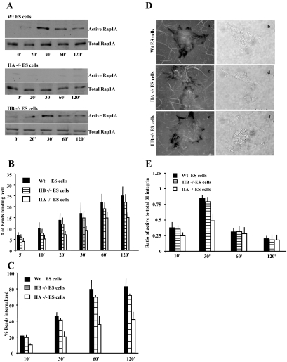
Figure 7.
(A) GST-RalGDS fusion protein was used to determine levels of GTP-bound Rap1 in the WT, NM II-A null cells, and NM II-B null ES cells after incubation with collagen-coated beads. (B) NM II-A null, NM II-B null, and WT ES cells were incubated on collagen bead-coated plates for varying times and washed to remove unbound beads. NM II-A null cells exhibit lower numbers of bound beads per cell compared with WT and NM II-B null cells. Data represent the mean ± SEM for three independent experiments, 25 cells per experiment. (C) NM II-A null, II-B null, and WT ES cells exhibit similar numbers of internalized collagen beads as a function of bound beads. (D) NM II-A null, II-B null, and WT ES cells plated on FITC-labeled collagen substrates for 4 h. NM II-B (e and f) and WT (a and b) cells show substantial substrate clearance (i.e., collagen uptake) compared with NM II-A null ES cells (c and d) as indicated by dark areas depleted of fluorescent collagen surrounding the cells (fluorescence images a, c, and e; phase images b, d, and f). (E) NM II-A null ES cells show reduced β1-integrin activation at 30 min compared with NM II-B and WT ES cells.

