Figure 1.
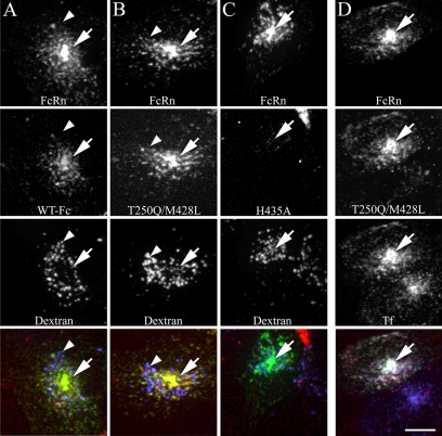
Fc colocalizes with human FcRn and transferrin in recycling compartments and is not present in lysosomes during short-term labeling. Cells expressing human GFP-FcRn were labeled for 20 min with 200 μg/ml TxR-Fc (4.4 μM) at pH 7.4 and then fixed. Cells were either prelabeled with Alexa 647-dextran as a lysosomal marker (50 μg/ml overnight dextran incubation chased to lysosomes for 1 h before addition of Fcs; A–C) or labeled simultaneously with 30 μg/ml Cy5-Tf (D). Projections were made of three-dimensional (3D) image volumes for each fluor and were merged as single images in the bottom row (green, GFP-FcRn; red, Fc; blue, dextran; yellow, colocalization of GFP-FcRn and FC; white (bottom right image only), three color colocalization of GFP-FcRn, Fc, and transferrin). WT Fc (A) and the T250Q/M428L mutant Fc (B) strongly colocalize with human FcRn with prominent fluorescence in the pericentriolar recycling compartment (arrows). Very little fluorescence is observed in the dextran lysosomal compartment (arrowheads). Fluorescent Fc endosomes are very dim in cells labeled with the H435A mutant (C). Cells labeled with Fc and transferrin show extensive colocalization between Fc, transferrin and FcRn (D). Note untransfected cell in D (bottom cell), which labels with transferrin but shows very little Fc internalization. All images from a given fluor were processed and contrast-adjusted in an identical manner, with the exception of the WT Fc images, which were contrast-stretched threefold relative to T250/M428L in order to visualize the overall dimmer labeling, and the H435A images, which were likewise stretched threefold for comparability to the WT Fc images. Scale bar, 10 μm.

