Figure 1.
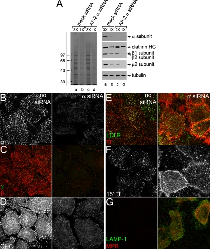
siRNA-mediated gene silencing of the AP-2 α subunit. (A) Lysates of HeLa SS6 cells untreated (lanes a and b) or treated with AP-2 α subunit siRNA (lanes c and d) were resolved by SDS-PAGE and either Coomassie stained or transferred to nitrocellulose. Sections of the blots were probed with anti-AP-2 α subunit mAb clone 8, anti-clathrin heavy chain (HC) mAb TD.1, anti-β1/β2 subunit mAb 100/1, anti-AP-2 μ2 antiserum, or anti-tubulin mAb E7, and only the relevant portion of each blot shown. The position of molecular mass standards (in kilodaltons) is indicated on the left. (B–D) HeLa SS6 cells untreated (left) or treated with AP-2 α subunit siRNA (right) were treated with 10 μg/ml brefeldin A for 15 min, permeabilized on ice for 1 min before fixation, and prepared for immunofluorescence using the anti-AP-2 α subunit mAb AP.6 (B), anti-AP-1 γ subunit mAb (C; green), anti-β1/β2 subunit antibody GD/2 (C; red), or anti-clathrin heavy chain mAb X22 (D). (E–G) HeLa SS6 cells untreated (left) or treated with AP-2 α subunit siRNA (right) were incubated on ice for 1 h with either anti-LDL receptor mAb C7 (E; green), Tf568 (E, red and F), anti-LAMP-1 mAb (G; green), or anti-MPR antibody (G; red) and fixed (E and G) or washed and warmed to 37°C for 15 min and fixed (F) followed by indirect immunofluorescence. Bar, 10 μm.

