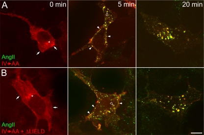
Figure 12.
Down-regulation of the type 1 angiotensin II receptor by both tdRFP-β-arrestin 1 (IV→AA) and β-arrestin (IV→AA+ΔLIELD). HEK293 cells stably expressing FLAG-tagged type I angiotensin II receptor were transiently transfected with tdRFP-β-arrestin 1 (IV→AA) (A), or tdRFP-β-arrestin 1 (IV→AA+ΔLIELD) (B). Cells were starved for 1 h in starvation medium and then stimulated with 100 nM angiotensin II conjugated to Alexa488 (AngII; green) for 0, 5, or 20 min. Cells were washed, fixed, and examined by confocal microscopy. Representative images of medial focal planes are shown. Before stimulation, β-arrestin is largely diffuse in the cytosol but also present at the plasma membrane (arrows) or at a juxtanuclear location (asterisks). After 5 min of stimulation, β-arrestin clusters with angiotensin II at the plasma membrane (closed arrowheads), and on endosomes (open arrowheads). At 20 min after stimulation, nearly all β-arrestin and angiotensin II colocalize on larger endosomes. Bar. 10 μm.

