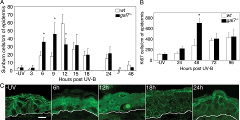
Figure 4.
In vivo UVB irradiation. The wt and galectin-7−/− adult female mice were depilated and irradiated with 2000 J/m2 UVB. (A) Apoptotic kinetics. Sections of unirradiated (−UV) or irradiated back skin samples were stained with hematoxylin and eosin, and the number of sunburn cells per centimeter of epidermis was determined. Three to six mice per genotype were used for each time point. Each bar represents the mean value ± SD. Statistical differences between wt and mutant animals are noted *p < 0.01. (B) Proliferative response. Sections of unirradiated (−UV) or irradiated back skin samples were labeled with anti-Ki67 Ab, and the number of positive cells per centimeter of epidermis was determined. Three to six mice per genotype were used for each time point. Each bar represents the mean value ± SD (*p < 0.01) (C) Confocal analysis of galectin-7 distribution. Representative sections of unirradiated (−UV) or irradiated wt back skin samples were stained with anti-galectin-7 Ab. White lines indicate the dermoepidermal junction. Bar, 10 μm.

