Figure 6.
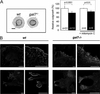
Galectin-7 in reepithelialization process ex vivo. (A) Picture of wt and galectin-7−/− newborn skin explants processed for keratin17 immunostaining after 7 d in culture. Dashed lines indicate the leading edge of keratinocyte outgrowths. Bar, 2 mm. Keratinocyte outgrowth area of wt (white bars) and galectin-7−/− (black bars) explants were measured either after 7 d in normal culture medium (bars on the left) or when a 2 h mitomycin-C treatment had been applied on the second day. n, number of animals used. (B) Subcellular distribution of cortactin in lamellopodia of migrating keratinocytes in wt and galectin-7−/− explants. On day 7, cultures were fixed, permeabilized and stained with anti-cortactin antibody. Nuclei were detected by Hoechst 33342 staining. Bar, 20 μm.

