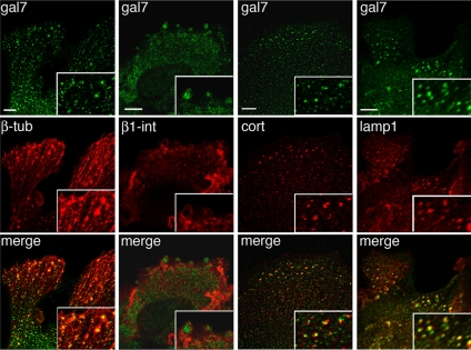
Figure 7.
Confocal analysis of galectin-7 distribution in leading edge keratinocytes. Double immunostainings with anti-galectin-7 Ab combined with anti-β-tubulin, anti-β1-integrin, anti-cortactin, or anti-Lamp1 Abs were performed on wt skin explants fixed after 7 d in culture. Pictures were taken in the focal plane of cell adhesion structures. Bar, 10 μm.

