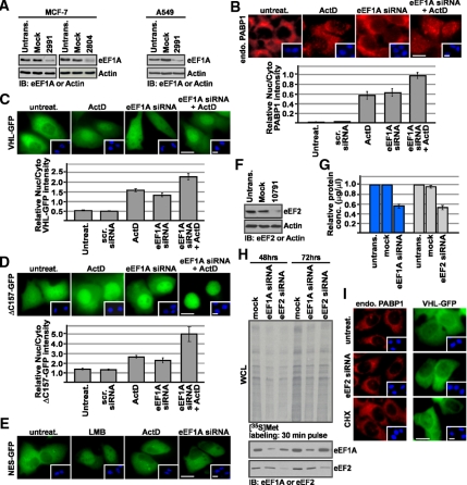
Figure 4.
siRNA-mediated silencing of eEF1A causes nuclear accumulation of TD-NEM–containing proteins. (A) eEF1A-specific siRNA reduces eEF1A protein level. MCF-7 and A549 cells were either mock-transfected or transfected with 100 nM eEF1A siRNA (2991 or 2804) for 72 h and then subjected to Western blot analysis using anti-eEF1A antibody. Actin was used as a loading control. (B) eEF1A knockdown and ActD treatment cause nuclear accumulation of endogenous PABP1. MCF-7 cells were transiently transfected with 100 nM control scrambled siRNA, 100 nM eEF1A siRNA (2991 or 2804), or untransfected for 72 h. Where indicated cells were transfected with 100 nM eEF1A siRNA for 72 h followed by treatment with 8 μM ActD for 3 h. Localization of endogenous PABP1 was determined by immunofluorescence using a PABP1-specific antibody. Scale bar, 10 μm. (C–E) eEF1A knockdown and ActD treatment results in nuclear accumulation of transiently expressed VHL-GFP and ΔC157-GFP, but does not affect GFP-NES. MCF-7 cells were cotransfected with 100 nM eEF1A siRNA (2991 or 2804) or control scrambled siRNA and VHL-GFP, ΔC157-GFP, or GFP-NES. Where indicated, cells were treated with 8 μM ActD or 10 μM LMB for 3 h after the 72-h incubation after transfection. Insets in B–E show Hoechst staining of DNA; scale bars, 10 μm. Graphs in B–D represent relative nuclear:cytoplasmic ratios of fluorescence intensity. (F–I) Nuclear accumulation of TD-NEM–containing proteins in the absence of eEF1A is not due to an overall decrease in protein translation. (F) A549 cells were either mock-transfected or transfected with 100 nM eEF2 siRNA (10791) for 72 h and then subjected to Western blot analysis using anti-eEF2 antibody. Actin was used as a loading control. (G) A549 cells were untransfected, mock-transfected, or transfected with eEF1A or eEF2 siRNA for 72 h. Total cellular protein levels were measured using a standard protein quantification method. The decrease in protein levels in mock- or siRNA-transfected cells was calculated relative to protein levels in untransfected cells. (H) A549 cells were mock-transfected or transfected with eEF1A or eEF2 siRNA for 48 and 72 h. At the indicated times, cells were pulse-labeled for 30 min with [35S]Met. Labeled whole cell lysates (WCL) were separated on a 10% SDS-polyacrylamide gel and transferred onto a PVDF membrane, and translational activity was visualized by autoradiography (top panel). The two bottom panels show immunoblots from the same membrane, using eEF1A and eEF2 antibodies. (I) For determining the effect of eEF2 silencing, cells were transfected with only 100 nM eEF2 siRNA or cotransfected with VHL-GFP and 100 nM eEF2 siRNA for 72 h. Endogenous PABP1 was detected by immunofluorescence using a PABP1 antibody. For determining the effect of cycloheximide, cells were either untransfected or transiently transfected with VHL-GFP followed by treatment with cycloheximide for 2 h. Insets, Hoechst staining of DNA; scale bars, 10 μm.

