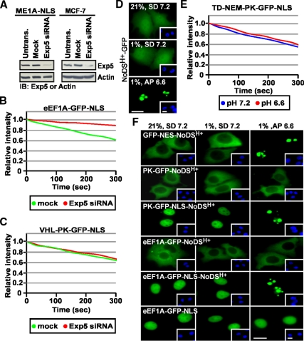
Figure 7.
The function of eEF1A in nuclear export of TD-NEM–containing proteins is Exp5-independent and exerted from the cytoplasmic side of the nuclear envelope. (A–C) Nuclear export by TD-NEM is independent of Exp5. (A) ME1A-NLS (MCF-7 cells stably expressing eEF1A-GFP-NLS) and MCF-7 cells were transfected with 100 nM Exp5 siRNA for 48 h and then lysed and subjected to Western blot analysis using an anti-Exp5 antibody. Actin was used as a loading control. ME1A-NLS cells (B) or MCF-7 cells transiently expressing VHL-PK-GFP-NLS (C) were mock-transfected or transfected with 100 nM Exp5 siRNA and incubated for 48 h after which nuclear export was analyzed by the live cell FLIP nuclear export assay, as previously described. Loss of nuclear fluorescence was plotted on a graph (B and C). (D–F) eEF1A is not targeted to the nucleolus by the NoDSH+. (D) MCF7 cells were cultured in standard media (SD) and transiently transfected to express the indicated GFP-tagged constructs. Cells were replenished with either fresh standard media (SD, pH 7.2) or acidification permissive media (AP, initial pH 7.2) that allows maximal extracellular acidification to pH 6.6 (see Materials and Methods). Cells either remained in normoxia (21% O2) or transferred to hypoxia (1% O2) for 18 h. Extracellular pH at the endpoint is indicated on each panel. (E) MCF-7 cells transiently expressing TD-NEM fused to PK-GFP-NLS were treated the same as in D. Cytoplasmic FLIP was performed to assess nuclear export activity. Loss of nuclear fluorescence was monitored and plotted on a graph. (F) MCF-7 cells transiently expressing the indicated constructs were incubated in the same conditions as in D. The localization of GFP-tagged fusion proteins was assessed by fluorescent microscopy. Insets in D and F show Hoechst staining of DNA; scale bars, 10 μm.

