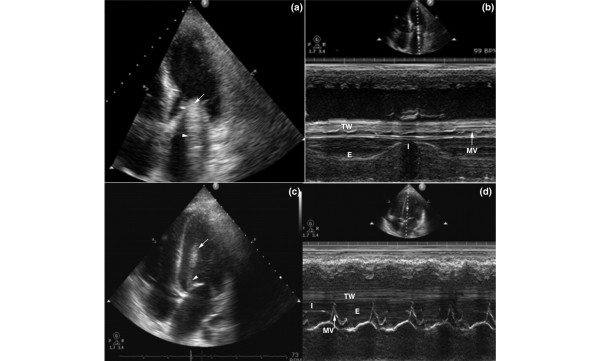
Figure 1.
Echocardiography depicting the artifact (arrows) E, expiration; I, inspiration; MV, mitral valve; TW, thoracic wall. (a,c) Apical four-chamber views and (b,d) M-mode of the artifact in the left ventricle. The immobile part (arrow) and the mobile one (arrowhead) may be observed. The respiratory variation that resembles the lung 'sinusoid sign' is either fully (panel b) or partially (panel d) visible.

