Figure 4.
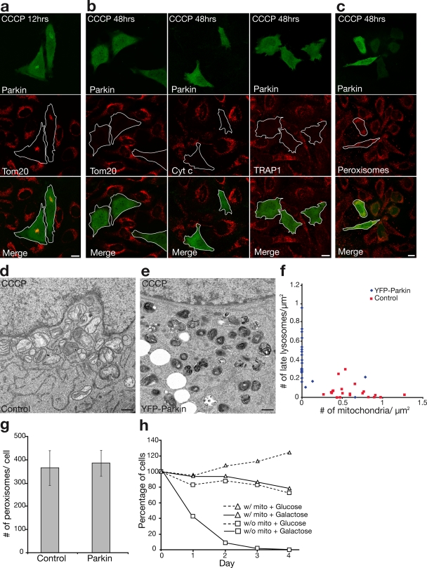
Selective mitochondrial elimination by Parkin under depolarizing conditions. (a and b) HeLa cells expressing YFP-Parkin (green) incubated for 12 h (a) or 48 h (b, left) with 10 μM CCCP. Cells were immunostained for Tom20 (red). Parkin-expressing HeLa cells display less mitochondrial mass compared with surrounding cells at 12 h and complete loss of mitochondria by 48 h. (b) Similar loss of mitochondria observed with anti–cytochrome c (red, middle) and anti-TRAP1 (red, right) antibodies. (c) No loss of peroxisomes immunostained for PMP70 (red) in YFP-Parkin–transfected cells relative to surrounding untransfected cells. Outlines demarcate the edges of cells expressing YFP-Parkin. Bars, 10 μm. (d–f) Electron microscopy of untransfected HeLa cells (d) or HeLa cells expressing YFP-Parkin (e) and treated with 10 μM CCCP for 48 h. Many mitochondria and few lysosomes were observed in control cells, and no mitochondria and many lysosomes were observed in YFP-Parkin–transfected cells. Bars, 500 nm. (f) The number of mitochondria and late lysosomes/μm2 of cytoplasm in 22 randomly selected cells per condition. (g) The number of PMP70-stained peroxisomes per cell in YFP-Parkin–transfected and untransfected cells (n = 5). Error bars indicate standard deviation of at least three replicates. (h) Control HeLa cells or HeLa cells transfected with YFP-Parkin treated with 10 μM CCCP for 72 h (day 0) and cultured in glucose or galactose media for 1–4 d. Cells were fixed and stained for Tom20 and Hoechst33342 (nuclei). Cells with nonapoptotic nuclei in a representative area of the slide on days 0–4 were counted and represented in the graph as a percentage of nonapoptotic cells on day 0 (≥160 cells per condition on day 0 in at least two experiments).

