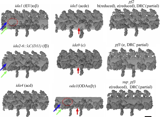
Figure 2.
Three-dimensional structure of mutants. Components missing in each mutant are indicated in parenthesis. In ida1, ida2-6∷λC(D11), ida5, ida9, and oda1 mutants, the locations of a dynein f dimer (blue and green) and dynein c (red) are indicated by arrows, whereas the location of the LC–IC complex of dynein f is encircled by red dotted circles. Bar, 20 nm.

