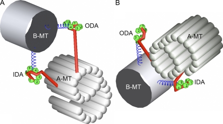
Figure 9.
Schematic diagram of the conformation of heavy chains in the inner and outer dynein arms. (A) The view from the B-microtubule (B-MT). (left) Proximal side. (right) Distal side. (B) The view from the A-microtubule (A-MT). (left) Distal side. (right) Proximal side. The AAA domains are marked. The assignment and the conformation of the six AAA domains (green balls) in each ring, the coiled-coil stalks (blue helices), and the N-terminal tails (red rods) are based on Burgess et al. (2004). The likely arrangement of the six AAA domains is indicated based on the two-dimensional averaging of dynein c (Burgess, S., personal communication). Combining this indexing and three-dimensional reconstruction of cytoplasmic dynein (Samso and Koonce, 2004), we speculate that the tail wraps around the AAA ring, as shown here. IDA, inner dynein arm; ODA, outer dynein arm.

