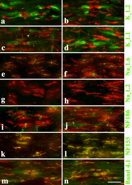
Figure 4.
Ion channels and structural components of the nodal region are appropriately localized in Claudin 11–null mice. Cryostat sections of adult optic nerve from 2% PFA (in 0.1 M sodium phosphate buffer, pH 7.4) perfused wild-type (a, c, e, g, i, k, and m) and Claudin 11–null mice (b, d, f, h, j, l, and n). Antibody labeling shows that the major ion channels and structural components of nodes, paranodes, and juxtaparanodes are appropriately localized (green). Anti-caspr antibodies are used to label paranodes in all panels (red). Bar, 10 μm.

