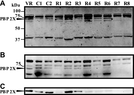
FIG. 3.
Binding of penicillin to GBS PBPs and immunodetection of PBP 2X. Membrane proteins were incubated with Bocillin FL, separated on a 10% (A) or a 5% (B) SDS-polyacrylamide gel, and detected by fluorography, as described in Materials and Methods. The molecular sizes of the protein standards (Precision Plus; Bio-Rad Laboratories, Hercules, CA) are provided on the left. (C) Membrane proteins separated on a 5% SDS-polyacrylamide gel were subjected to Western blotting with anti-PBP 2X antibody as the primary antibody, as described in Materials and Methods. Arrows indicate the position of PBP 2X. VR, S. agalactiae 2603 V/R.

