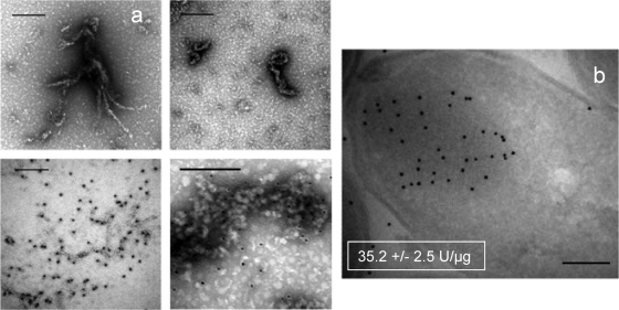
FIG. 2.
(a) Transmission electron microscopy analysis of fast protein liquid chromatography-purified GFP from peak 1, showing fibril-like and particulate microaggregates (top). At the bottom, the results of immunodetection of GFP on the same samples are shown. (b) Immunolabeling of GFP in inclusion bodies, on cryosections of GFP-producing E. coli cells. In the inset figures, the specific fluorescence values of GFP determined on isolated inclusion bodies are indicated. Bars, 0.2 μm.

