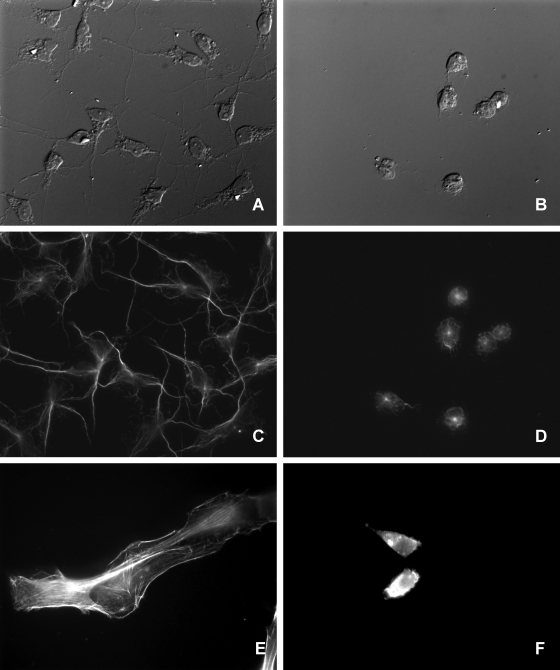
FIG. 3.
ECP alteration of cell morphology. Nonconfluent cell cultures were obtained by seeding coverslips with 65,000 Bge or 40,000 NIH cells in 300 μl of medium in 24-well plates and were grown overnight under the respective culture conditions. ECPs were added to the cells at a final protein concentration of 10 μg ml−1, and cells were further incubated for 6 h. (A and B) DIC microscopy images. ECP-treated Bge cells (B) showed cytoplasmic retraction and cell rounding compared to untreated Bge cells (A). (C to F) Fluorescence microscopy images. (C and D) The immunostaining of β-tubulin on Bge cells showed the retraction of the microtubule cytoskeletons of ECP-treated cells (D) compared to those of untreated cells (C). (E and F) NIH cells. (F) Actin labeling showed the disruption of the actin cytoskeletons of ECP-treated cells. (E) Untreated cells.

