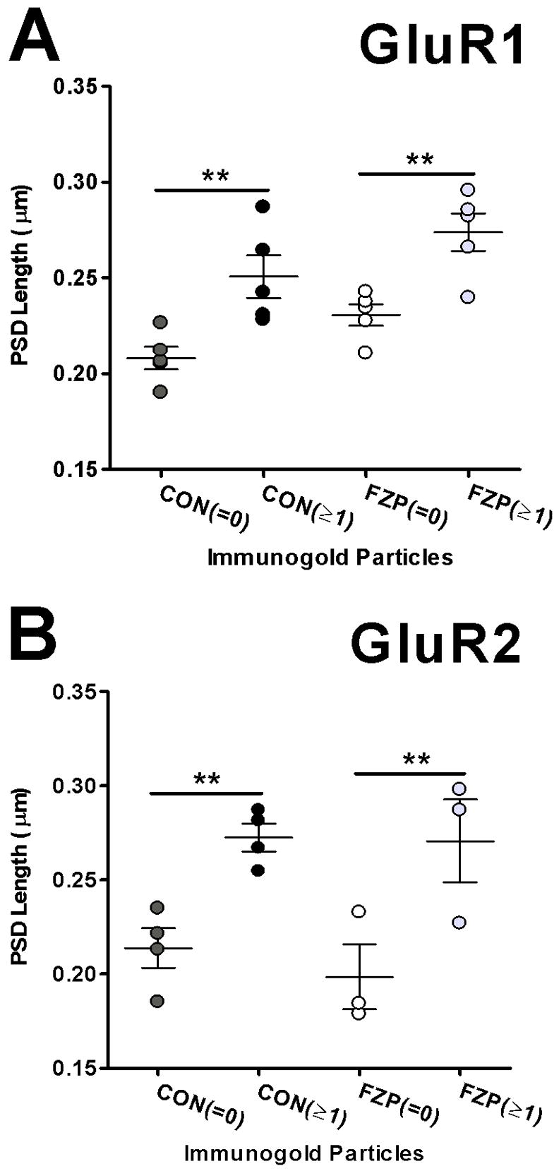
Figure 6.

Analysis of AMPA-immunonegative and immunopositive synapse size. (A) Analysis of PSD lengths in GluR1-labeled sections show that AMPAR immunonegative synapses (=0) are smaller than immunopositive synapses (≥1) in both control and FZP-withdrawn tissues (n=5 rats/group). (B) PSD lengths of immunonegative synapses from sections reacted with the GluR2 antibody were also significantly smaller than immunopositive synapses (**p<0.01).
