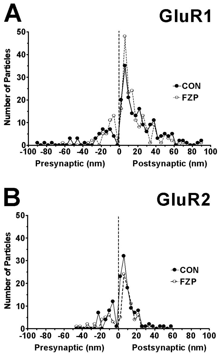
Figure 8.

Spatial distribution of all (A) GluR1, (B) GluR2 immunogold particles found within 100 nm of the postsynaptic membrane on either the extracellular or cytoplasmic side. Asymmetric synapses with clearly visible and well-defined PSDs and synaptic clefts were digitally imaged at a final magnification of X 36,000. The distances between centers of each 10 nm gold particle to the outer leaflet of postsynaptic membrane were measured and grouped into 4 nm wide bins (0 on the abscissa represents external face of postsynaptic membrane). Negative values indicate gold particles located at a distance from the postsynaptic membrane and in the direction of the presynaptic bouton and synaptic cleft (average width ~20 nm). Positive values indicate immunogold labeling on the cytoplasmic side. GluR1 and GluR2-immunogold labeling for both subunit antibodies peaked 6 nm inside the postsynaptic membrane (resolution ±2 nm) with GluR1 labeling extending more distally towards the cytoplasmic side than GluR2 labeling. The majority of both GluR1 and GluR2 immunolabeling was located within the PSD region (width ~45 nm). Very few immunogold particles were detected extracellularly, beyond the synaptic cleft (values <−20 nm). Thus, both the GluR1 and GluR2 immunolabeling analyzed was very tightly related to the extent of the PSD. Data was obtained from 67 (CON) and 56 (FZP) synapses in GluR1-labeled tissues and from 44 (CON) and 40 (FZP) synapses in GluR2-labeled tissues.
