
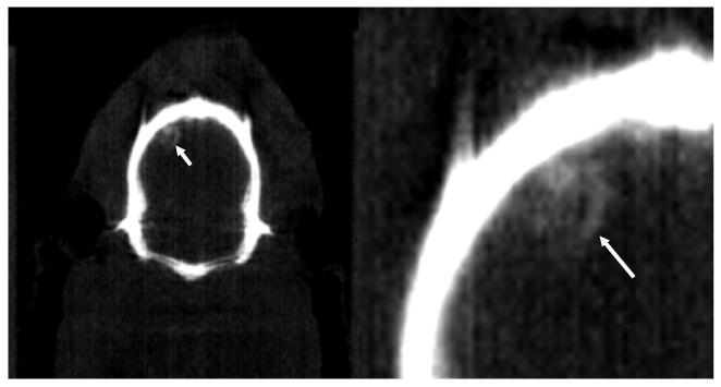
Figure 3.
A coronal view (left) of the presence of arterial blood in a noncontrast CT image (CT window: 0–1500 HU) indicated by a white arrow with a close-up view (right). The volume of femoral arterial blood injected was less than 0.1 ml.
Official websites use .gov
A
.gov website belongs to an official
government organization in the United States.
Secure .gov websites use HTTPS
A lock (
) or https:// means you've safely
connected to the .gov website. Share sensitive
information only on official, secure websites.
A coronal view (left) of the presence of arterial blood in a noncontrast CT image (CT window: 0–1500 HU) indicated by a white arrow with a close-up view (right). The volume of femoral arterial blood injected was less than 0.1 ml.