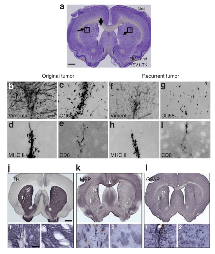
Figure 6. Neuropathological correlates in long-term survivors after glioblastoma multiforme rechallenge; minimal long-term side-effects.
The animals described in Figure 5a, treated with Ad-Flt3L and Ad-TK and surviving the primary and recurrent tumors, were killed 240 days after the initial tumor implantation and were subjected to immunohistochemical evaluation for neuropathology and the presence of immune cellular infiltrates in both hemispheres of the brain. Representative brain sections are shown. (a) Nissl staining reveals scar tissue adjacent to the injection site in both hemispheres and an enlarged ventricle ipsilateral to the site of the original tumor implantation. (b,f) Vimentin staining and morphological analysis of immunoreactive cells suggests the presence of activated astrocytes at both injection sites. Immunoreactivity for (c,g) macrophages, (d,h) major histocompatibility class II (MHC II) + immunoreactive cells, and (e,i) CD8 + T cells reveals low levels of infiltration of immune cells both at the original and recurrent tumor injection sites. (j,k) Immunoreactivity with markers for tyrosine hydroxylase (TH) and myelin basic protein (MBP) reveals slight tissue disruption in close proximity to the injection site, but otherwise normal expression patterns throughout the striatum. Images showing disruptions to (j1) nerve terminals and (k1) axon bundles are limited to the injection sites. Images from an area of the striatum distant from the injection site show (j2) normal TH distribution and (k2) MBP-coated axons. (l1) Increased glial fibrillary acidic protein (GFAP) immunoreactivity is localized to areas close to both injection sites, indicating the presence of activated astrocytes. (l2) Normal GFAP immunoreactivity is observed at an area of the striatum distant from the injection site. Ad, adenovirus; Flt3L, Fms-like tyrosine kinase 3 ligand; TK, thymidine kinase.

