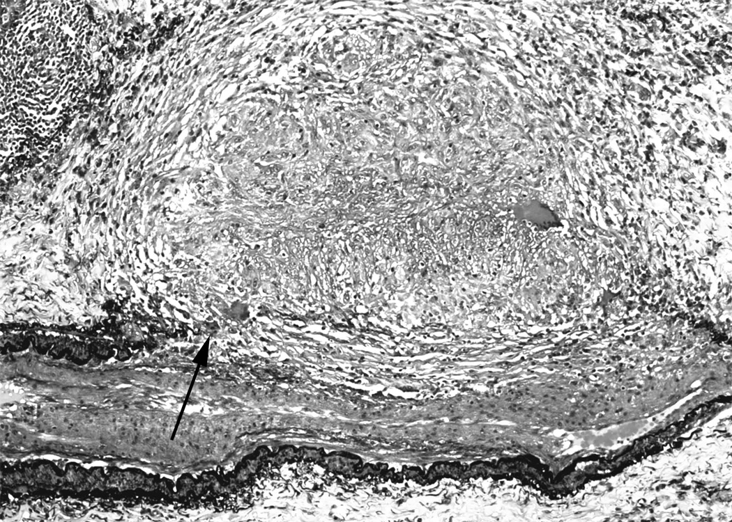
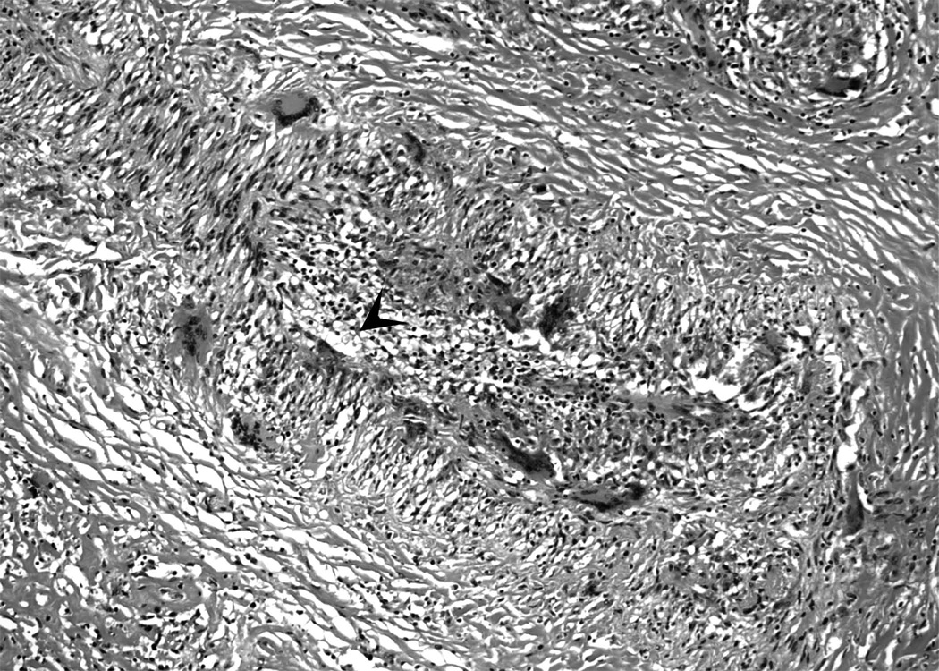
Figure 2. Granulomatous arteritis in sarcoidosis.
2a. Movat pentachrome stain showing granulomas obliterating the internal and external elastic laminae and smooth muscle (arrow) of medium size artery. There is secondary intimal fibroplasia seen in the center of the artery. (Movat pentachrome).
2b. In some cases, only giant cells and mononuclear cell infiltrates may be seen in sarcoidosis-associated arteritis. Arrowhead indicates the vascular lumen. Numerous giant cells may be seen in the vessel wall (Hematoxylin and eosin).


