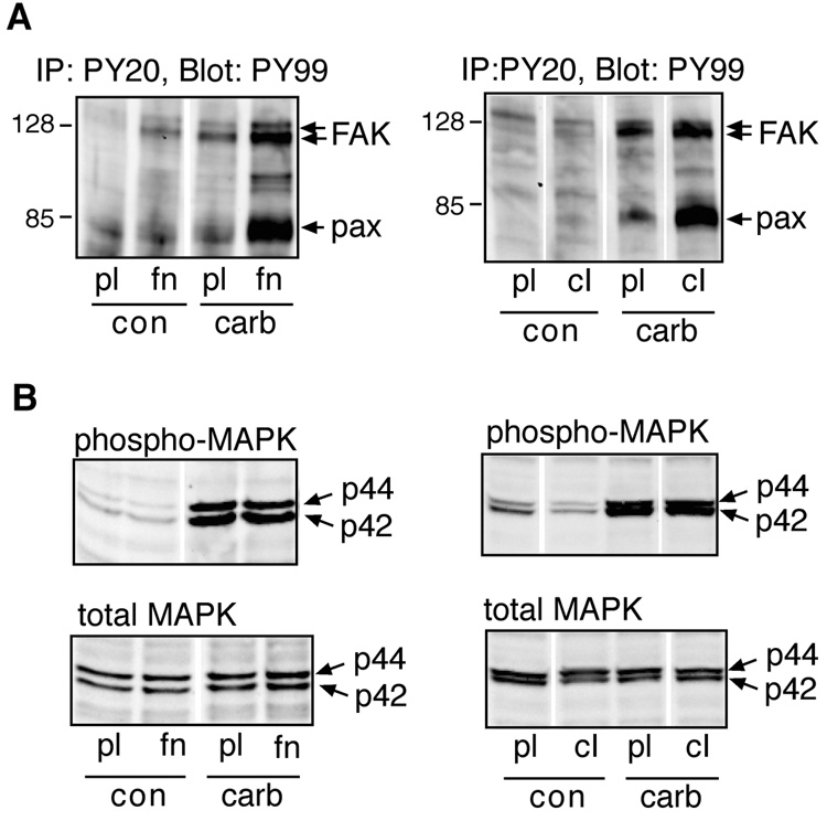
Fig. 1.
Effect of ECM proteins on M3 receptor-evoked tyrosine phosphorylation and MAPK activation. Serum-deprived HEK-M3 cells were placed in suspension, and replated onto culture dishes coated with polylysine (pl), fibronectin (fn), or collagen type I (cI). The cells were allowed to adhere for 2.5 hours, then stimulated with control medium (con), or medium containing 100 µM carbachol (carb), for 20 minutes. (A) Tyrosine phosphorylated proteins were immunoprecipitated (IP) with anti-phosphotyrosine antibodies (clone PY20), resolved by SDS-PAGE, and immunoblotted with peroxidase-linked anti-phosphotyrosine antibodies (clone PY99). Arrows indicated paxillin (pax) and focal adhesion kinase (FAK). Note that FAK appears as a doublet in this cell line [Slack, 1998]. (B) Total MAPK and phospho-MAPK were detected by immunoblot analysis of cell lysates.

