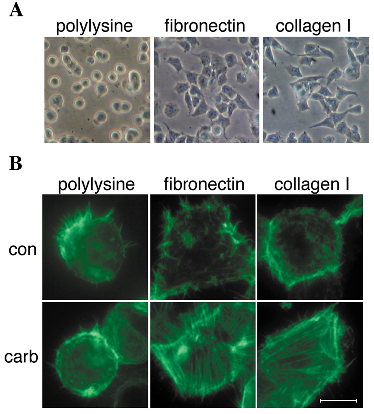
Fig. 2.
Cell spreading and M3 receptor-evoked stress fiber formation is promoted by fibronectin and collagen type I. HEK-M3 cells were serum-deprived overnight, placed in suspension and replated onto dishes coated with polylysine, fibronectin, or collagen type I. (A) Cells were allowed to adhere for 1 hour, and photographed under phase-contrast using a 10X objective. (B) Cells replated on coverslips coated with polylysine, fibronectin, or collagen type I were allowed to adhere for 2.5 hours, then treated with control medium (con), or medium containing 100 µM carbachol (carb), for 20 minutes. Cells were fixed and permeabilized, and immunofluorescence labeling was carried out with Alexa Fluor 488-conjugated phalloidin to detect F-actin. Bar, 10 µM.

