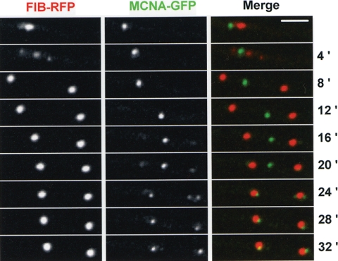
FIG. 8.
Segregation of MCNA bodies occurs through a similar mechanism to nucleolar segregation but is completed later in the cell cycle. Nuclear division of the nucleolus following Fib-RFP and MCNA bodies, shown using MCNA-GFP. At 4 min Fib-RFP is at a mid-stage of segregation, displaying a central cytoplasmic parental nucleolus flanked on either side by newly forming daughter nucleoli. By 8 min the nucleolus has completed its segregation to daughter nuclei but the MCNA body remains intact within the cytoplasm. At 16 min the parental MCNA body begins to diminish as new MCNA bodies appear in daughter nuclei next to the nucleoli. As the cell cycle continues the parental MCNA body disappears and new MCNA bodies are formed in daughter nuclei. Bar, ∼5 μm.

