FIG. 3.
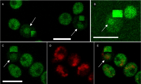
Gal3p-GFP localizes to the nucleus and cytoplasm. Gal3p-GFP was constructed as part of the whole-genome GFP-tagging project (13). (A) Removal of the cytoplasmic signal by bleaching shows nuclear-localized Gal3p-GFP as indicated by the arrows. (B) Gal3p is observed in the nucleus shortly after transfer to a galactose-containing medium. Gal3p-GFP was grown overnight in raffinose and transferred to fresh medium containing galactose and incubated for 45 min. The arrow indicates the nuclear-localized Gal3p. (C, D, E) Cytoplasmic bleaching reveals nuclear Gal3p-GFP. Gal3p-GFP is shown in panel C, DAPI in panel D, and the merged image in panel E. The persistence of nuclear GFP signal in the bleached cell is shown by the arrow in panels C and E. Bars = 5 μm.

