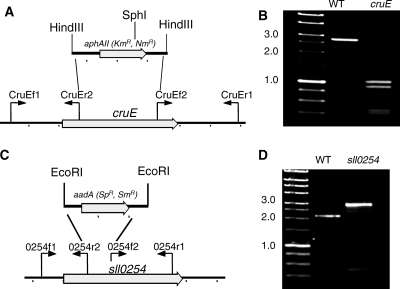
FIG. 3.
Restriction maps and PCR verification of cruE mutants of two cyanobacteria. (A and C) Restriction maps showing construction of disruption mutations of the cruE gene of Synechococcus sp. strain PCC 7002 (A) and the sll0254/cruE gene of Synechocystis sp. strain PCC 6803 (C). (B and D) Agarose gel electrophoresis of PCR amplicons from the wild-type (WT) and cruE mutant strains of Synechococcus sp. strain PCC 7002 (B) and wild-type and sll0254/cruE mutant strains of Synechocystis sp. strain PCC 6803 (D). In panel B the PCR amplicons were both digested with SphI to demonstrate the difference, because the inserted drug resistance cartridge was nearly identical in size to the DNA fragment that had been deleted. The data show that both mutant strains are completely segregated. Size markers are indicated at the left of panels B and D, and selected sizes in kb are indicated.

