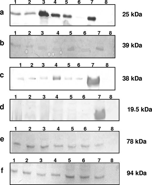
FIG. 3.
Western immunoblot analyses of EspA (a), EspD (b), EspB (c), bundle-forming pilus (d), Tir (e), and intimin (f) in O125ac:H6 atypical EPEC strains. Secreted (a to c) or whole-cell (d to f) proteins were separated on 10% sodium dodecyl sulfate-polyacrylamide gels, and the transferred nitrocellulose membranes were immunodetected with the corresponding polyclonal antiserum. Lanes: 1, EC292/84; 2, 1794/80; 3, CB1924; 4, CB5304; 5, CB3114; 6, CB3338; 7, positive controls (E2348/69 [a to d and f] or EDL933 [e]); 8, negative controls (UMD872 [a], UMD870 [b], UMD864 [c], JPN15 [d], or E. coli DH5α [e and f]). The apparent molecular masses are indicated on the right.

