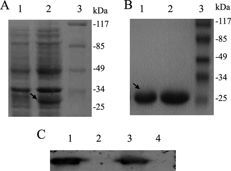
FIG. 1.

P protein expression, purification, and antibody preparation. (A) SDS-PAGE of the induction and expression of His-P protein in E. coli BL21. The gels were stained with Coomassie brilliant blue R-250. Lane 1, proteins expressed in BL21 cells transformed with pET-P without IPTG induction; lane 2, proteins expressed in BL21 cells transformed with pET-P and induced with 1 mM IPTG; lane 3, prestained protein markers 0431. (B) SDS-PAGE of purified His-P protein. Lanes 1 and 2, purified His-P protein; lane 3, prestained protein markers 0431. (C) Western blot of antibody-recognized P protein. Lane 1, proteins prepared from OL BDV-infected (OL/BDV) cells detected by Western blotting using polyclonal antibody to P protein; lane 2, protein preparation of uninfected (OL) cells detected by Western blotting using polyclonal antibody to P protein; lane 3, protein preparation of 293T cells transfected with pcDNA-P detected by Western blotting using polyclonal antibody to P protein; lane 4, protein preparation of 293T cells transfected with pCNDA-3 detected by Western blotting using polyclonal antibody to P protein.
