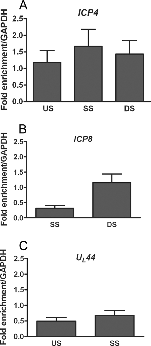
FIG. 1.
ChIP analysis of the fraction of HSV-1 DNA associated with histone H3. Total cell extracts were prepared at 3 hpi from HeLa cells infected with HSV-1 at an MOI of 0.1 PFU/cell. ChIP was carried out with an antibody specific to the C terminus of histone H3. The fraction of DNA immunoprecipitated compared to the input value was determined by real-time PCR, with the fraction immunoprecipitated by a nonspecific antibody subtracted, and is expressed as the fold enrichment over the fraction of GAPDH DNA immunoprecipitated. Primers were specific for regions of ICP4 (A), ICP8 (B), and UL44 (C) (see Table 1 for primer locations and sequences). Abbreviations: US, upstream of the transcriptional start site; SS, start site; DS, downstream of the transcriptional start site. The means and standard errors of the means of at least five independent experiments are shown.

