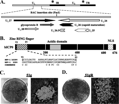
FIG. 1.
BHV-1 BAC organization and analysis of wt BHV-1 BAC. (A) BHV-1 genomic organization, showing the intergenic region between the UL27/UL28 and UL26/UL26.5/UL25/UL24 genes and the relative transcription directions/orientations of these genes. The site of the BAC insertion (PacI site) is shown. The position of the bICP0 gene located in the inverted repeat (IR) and the terminal repeat (TR) is denoted by the asterisk. (B) The bICP0 protein is 676 aa long and contains several important functional domains. The bICP0 zinc RING finger (aa 13 to 51) is similar to the zinc RING finger present in the HSV-1 ICP0 protein. Positions of conserved C residues in a consensus C3HC4 zinc RING finger are shown. The locations of the acidic domain and the nuclear localization signal (NLS) within bICP0 are also denoted. The numbers below the diagram denote amino acid sequence numbers of bICP0. Mutagenesis of aa 13 (cysteine to glycine) and aa 51 (cysteine to alanine) (underlined) impairs the ability of bICP0 to activate transcription in transient-transfection assays (34), and plasmids containing these mutated cysteines do not inhibit IFN-dependent transcription (28, 63). (C and D) CRIB cells were infected with 51g (C) or 51gR (D) at an MOI of 0.1. After incubation at 37°C for 1 h, cells were rinsed three times with calcium magnesium-free PBS and replaced with fresh media. Plaques were stained with crystal violet at 72 h after infection for 51g or at 36 h after infection for 51gR. Images were taken at a ×10 (left panels) or a ×100 (right panels) magnification using a Leica DM IRB microscope.

