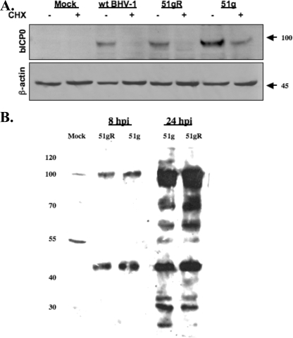
FIG. 2.
Western blot analysis of bICP0 and viral proteins in productively infected cells. (A) CRIB cells were mock infected or infected with the 51g mutant, 51gR, or wt BHV-1 at an MOI of 1.0. At 6 h after infection, some cultures were treated with 100 μg/ml cycloheximide (Sigma) (CHX; + lanes) to inhibit protein synthesis. Two hours later (8 h after infection), cells were lysed by treatment with NP-40 and sonication and then processed for Western blot analysis as described in Materials and Methods. Following SDS-PAGE (8%), immunoblotting was performed with a polyclonal antibody generated against the N-terminal 361 aa of bICP0 (1:500). The bICP0 protein migrates at approximately 97 kDa, and β-actin migrates at approximately 45 kDa (marked at right). For each lane, 50 μg protein was added. (B) CRIB cells were infected with the designated strains of BHV-1 for 8 or 24 h postinfection (hpi). After cells were lysed, proteins (50 μg/lane) were electrophoresed by 8% SDS-PAGE. Immunoblotting was performed using an anti-BHV-1 virion antibody that was commercially available from VMRD (Pullman, WA). Markers (in kilodaltons) are shown at left.

