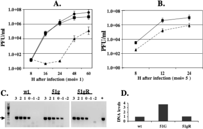
FIG. 3.
Growth of the 51g mutant, 51gR, or wt BHV-1 in CRIB cells. CRIB cells were infected with the 51g mutant, 51gR, or wt BHV-1 at an MOI of 1.0 (A) or 5.0 (B). Error bars show the standard errors of the results of three independent studies. (C) Viral DNA was prepared from 107 PFU of virus stocks that were obtained from CRIB cells infected with wt BHV-1, the 51g mutant virus, or 51gR. Tenfold dilutions of viral DNA were amplified using gB-specific primers (forward, 5′-GTGGTGGCCTTTGACCGCGAC-3′; reverse, 5′-GCTCCGGCGAGTAGCTGGTGT-3′). Amplified products were electrophoresed on a 1% agarose gel, and the ethidium bromide gel was photographed. The values above the lanes refer to the log of the original MOI of the respective virus stocks. The arrow denotes the position of the gB-specific amplified product. The “+” lane denotes a positive control. The lowest dilution of virus stocks that yielded a visible band was quantified using an RX biomolecular imager (Bio-Rad), and the arbitrary DNA levels are shown (D). These results are representative of comparing two different stocks of the respective viruses.

