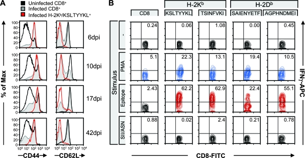
FIG. 6.
Epitope-positive CD8 T cells are activated. (A) Splenocytes from uninfected and infected mice were surface stained with CD44 and CD62L at 6, 10, 17, and 42 dpi. Representative stains are shown for each time point for the total CD8 T-cell population and for one of the epitope-positive populations (H-2Kb/KSLTYYKL). (B) In vitro stimulation of splenocytes from MHV-68-infected mice with no peptide (−), phorbol ester (phorbol myristate acetate [PMA]) and ionomycin, selected MHV-68 epitopes (the early KSLTYYKL/H-2Kb epitope, the late TSINFVKI/H-2Kb epitope, the early SAIENYETF/H-2Db epitope, and the late AGPHNDMEI/H-2Db epitope), or the known H-2Kb binding peptide SIINFEKL (SII) or known H-2Db binding peptide ASNENMDAM (ASN) demonstrates that CD8 T cells produce IFN-γ in an epitope-specific fashion. The data shown are representative of independent experiments. APC, allophycocyanin; FITC, fluorescein isothiocyanate.

