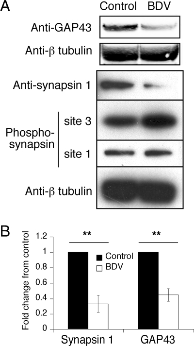
FIG. 3.
Expression of GAP-43 and synapsin 1 is impaired following BDV infection. (A) Western blot analysis of neuronal extracts from control and BDV-infected cultures using antibodies specific for GAP-43, total synapsin 1, phosphosynapsin 1 (site 3, specific for CaMK II, and site 1, specific for PKA and CaMK I), and beta-tubulin for normalization. Results are representative of four independent experiments. (B) Quantification of the Western blot signals using the Odyssey imager. Due to the intrinsic variability between neuronal cultures, results were expressed as their change compared to control neurons that were arbitrarily set to 1 in all cases. **, P < 0.05 by paired t test.

