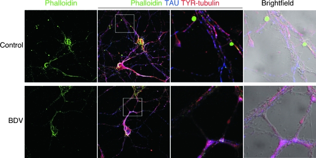
FIG. 4.
BDV infection interferes with localization of F actin at contact nodes between neurites. Shown is confocal microscopic imaging of control (top) and BDV-infected (bottom) cortical neurons triply stained for F actin using phalloidin-fluorescein isothiocyanate (FITC) (green), the neuronal marker Tau (blue), and the tyrosinated form of polymerized tubulin (TYR) to stain microtubule structures (red). Original magnification, ×630. An enlarged view of the highlighted white square of the triple-merge image is displayed on the two right pictures (fluorescence and bright field). At this higher magnification, note that the punctuated phalloidin staining colocalizes with contact nodes between neurites in control neurons and is lost following BDV infection.

