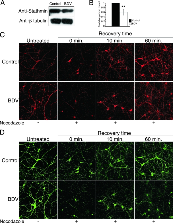
FIG. 5.
Infection with BDV delays recovery of the microtubule network following depolymerization with nocodazole. (A) BDV impairs stathmin expression. Shown is a Western blot analysis of neuronal extracts from control and BDV-infected cultures using antibodies specific for the microtubule-associated protein stathmin and beta-tubulin for normalization. Data are representative of two independent experiments. (B) Quantification of the Western blot signals using the Odyssey imager. **, P < 0.05 by paired t test. (C and D) Analysis of the microtubule network recovery kinetics following nocodazole-induced depolymerization. Neurons were treated (+) or not (−) with nocodazole and processed for immunofluorescence analysis at different times following washout of nocodazole. (C) Confocal microscopy imaging of staining using an antibody specific for the stable acetylated form of tubulin (ACE) (red). (D) Confocal microscopy imaging of staining using an antibody specific for the microtubule-associated protein Tau (green). Note the incomplete recolonization of the microtubule network by Tau even after 1 h of recovery time. Original magnification, ×400.

