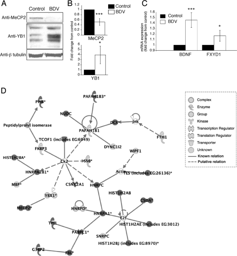
FIG. 6.
Infection with BDV modifies the expression pattern of the MeCP2 and YB1 proteins as well as that of MeCP2 target genes. (A) Western blot analysis of neuronal extracts from control and BDV-infected cultures using antibodies specific for MeCP2, YB1, and beta-tubulin for normalization. Results are representative of four to six independent experiments. (B) Quantification of the signals using the Odyssey imager. Due to the intrinsic variability between neuronal cultures, results were expressed as their change compared to control neurons that were arbitrarily set to 1 in all cases. ***, P < 0.0; *, P < 0.1 (by paired t test). (C) Real-time quantitative reverse transcription-PCR analysis of the expression of MeCP2 target genes. Levels of BDNF and phospholemman (FXYD1) transcripts were determined using total RNA extracted from control and BDV-infected neurons. Transcript levels were normalized to HPRT. Results are displayed as means ± standard errors of the means for six independent experiments. ***, P < 0.01; *, P < 0.1 (by paired t test). (D) Representative example of the signaling network/function analyses performed using IPA. The list of identified proteins was analyzed using IPA tools as described in the text. The network shown in the figure had the highest score (score of 49) and included 30 focus proteins. The legend for each node shape and arrow is indicated at the bottom. Focus proteins were identified mainly in BDV-infected neuronal extracts, whereas others were found mainly in control neurons.

